Where Do We Stand on Inner Ear Imaging Technology?

Samir

Manager
Author
Staff
Benefactor
Jan 3, 2017
1,138
Sweden
Tinnitus Since
12/2016
Cause of Tinnitus
Accoustic trauma
Hello!

I am researching available options for imaging the inner ear for the purpose of studying and diagnosing inner ear pathologies, the cochlea in particular. It seems that existing technology is very limited in this respect. Unfortunately, the only way to study the human cochlea up-close is to use the cochlea of a dead person, or have the cochlea of a living person removed.

There is a technique where sound is used to test the cochlear function. This is based on Otoacoustic emissions (OAE) which were predicted by Thomas Gold in 1948 and demonstrated experimentally by David Kemp in 1978. This is probably the best technique we have right now for measuring inner ear health. However, I have read that this type of test is often inaccurate and the results are difficult to interpret. So are the results of this type of tests any more objective than simple audiograms? Have we not come any further since 1978?

It would be much better to be able to see healthy (or damaged) hair cells, rather than to hear them! Imagine having something like FMRI for the cochlea! Wouldn't that be something? Being able to peek inside the tiny cochlea of a living person non-invasively and having high resolution live view images displayed on a monitor, and seeing the vestibular, tectorial, the basilar membrane, and of course the stereocilia of hair cells in action? Is that kind of technology too much science fiction at the moment?

This may not give us a cure for tinnitus. But just imagine how much we could learn about the inner ear with a technology like that! A technology like that would give us the tool to answer many questions concerning inner ear pathologies and hearing disorders.
 
Hey Samir,

That is a great question!

I think the other key ingredient to learning about tinnitus is to unequivocally understand how the brain works, including all 100 billion neurons. In programming terms, if you understand how the code works, you can change it!
For example, imagine being able to give a blind person the gift of site when we identify the area of the brain that controls vision and fully understand the logic that lets us see. Or give a deaf person the gift of hearing?

I honestly don't think this is science fiction. Today, you have very smart people working on these problems and in my view, it is only a matter of time before breathtaking results are revealed.
They are definitely working on that very issue. The US government started the BRAIN Initiative 3 years ago and just a few months ago got a big surge of funding. Hopefully something comes of it within our lifetimes.

https://www.braininitiative.nih.gov/
 
They are definitely working on that very issue. The US government started the BRAIN Initiative 3 years ago and just a few months ago got a big surge of funding. Hopefully something comes of it within our lifetimes.

https://www.braininitiative.nih.gov/

Let's not forget the European equivalent of this initiative, called Human Brain Project (HBP).

https://www.humanbrainproject.eu/

I wonder why they decided to do parallel projects... wouldn't it be better to put their efforts and resources together?

The Human Genome Project was started in 1990 and it ended in 2003. With the current state of science and technology I think we might see the completion of BRAIN and HBP within 10 years. Had they put their efforts and resources together (Europe and USA) we might have seen a completion within 7 years, by the end of 2020. These projects may still be completed by the end of 2020 as it is, but I believe that mapping the brain is a much tougher problem than mapping the human genome. You also have that double work that everyone is doing. It seems wasteful to me, both in term of time and resources.
 
At the heart of the BRAIN and HBP is multi-photon microsocopy.


Each and every one of you can also help map the brain by playing the Eyewire game by MIT.
 
Multi-photon microscopy has also been used to study the mouse cochlea:
http://biomedicaloptics.spiedigitallibrary.org/article.aspx?articleid=1392727

For comparison, see these images of the organ of Corti of a mouse. All three images are taken through the round window at the same magnification. From left to right you see TPEF (two photon excitation fluorescence) microscope image, OPEF (one photon excitation fluorescence) microscope image, and wide-field transmission microscope image. Anyone can guess which one shows the most details.

jbo_18_3_031104_f003.png


Note that these are in situ images, meaning that the images were taken on site. In other words, the cochlea has not been removed for the purpose of imaging.

Abstract from a restricted access publication from the same lab (Molecular Neuro-Otology and Biotechnology Laboratory at Harvard):

Intracochlear imaging is of great interest clinically because cochlea is the central organ of hearing. However, intracochlear imaging is technologically challenging due to the cochlea's small size and encasement in bone.

The state-of- the-art imaging techniques are not adequate for high resolution cellular imaging to establish diagnosis without destroying the cochlea. We report in situ imaging of intact mouse cochlea using endogenous two-photon excitation fluorescence (TPEF) as the contrast mechanism. TPEF eliminates the need for exogenous labeling and eradicating the staining-induced artifacts. We used a natural, membranous opening into the cochlea, the round window, as the optical access to reach the organ of Corti, requiring no additional slicing or opening.

Our approach provides the maximum non-invasiveness in the imaging process. TPEF exhibits strong contrast allowing deep imaging of mouse cochlea with cellular and even subcellular resolution. Inner hair cell, outer hair cell and supporting cell are clearly identifiable in TPEF images. Distinct morphological differences are observed between healthy and noise-exposed cochleae, allowing detection of specific, noise-induced pathologic changes.

The TPEF images taken through the round window are correlated with the whole mount sections, verifying their reliability. Compared with one-photon excitation fluorescence (OPEF) confocal microscope and wide-field transmission microscope images taken under the same magnification and resolution, TPEF images demonstrate clear advantages in terms of sharpness, signal to noise ratio and contrast. These capabilities provide a working foundation for microendoscopy-based clinical diagnostics of sensorineural hearing loss.

This is all very promising technology. Each new discovery and development builds on the work of those that came before us. This would not have been possible without the GFP (green fluorescent protein) for which Osamu Shimomura, Martin Chalfie and Roger Y. Tsien earned the Nobel prize in chemistry in 2008.

Speaking of Nobel prize winners, let's not forget about Sir John B. Gurdon and Shinya Yamanaka who won the 2012 Nobel prize in medicine for "the discovery that mature cells can be reprogrammed to become pluripotent". This will certainly be very useful if we decide to use stem cells to regenerate inner ear hair cells.
 
There has also been some interesting work done by a team of researchers from Texas A&M University and Stanford University using optical coherence tomography (OCT). This is an imaging technology widely used by opticians. The research team has re-purposed this technology for studying the inner ear. They have used this technology to take in vivo images of the mouse cochleae.

https://www.ecnmag.com/news/2014/07/high-res-images-inner-ear-could-lead-new-hearing-loss-therapies

https://www.ncbi.nlm.nih.gov/pubmed/24448302

In the 2014 article linked above there is a mention of a working prototype device. Does anyone have any more news on the progress of this technology? The fact that they already had a working prototype back then seems very promising.
 
Besides the more conventional OCT, there is also a new development of this technology called micro OCT (µOCT). Have a look at the images below. Isn't that excellent level of detailed information?

srep33288-f2.jpg


These are in situ intracochlear images of a guinea pig. These images were captured at the same Harvard laboratory where the TPEF images above were captured. The full article can be found below.

Micro-optical coherence tomography of the mammalian cochlea:
http://www.nature.com/articles/srep33288

This is great! It means that at least one laboratory in the world is actively looking for a way to image the internals of the human cochlea.

These are great results! The technology is obviously here and it's working! It makes me wonder why this technology is not already being used in clinics? Doctors may tell you that you have sensorineural hearing loss. But how do they know? Based on an audiogram? When you ask them if it's possible to have a look inside the cochlea to see what's damaged, the answer is always No. It's impossible they say! It's obviously not impossible.
 
I wonder why they decided to do parallel projects... wouldn't it be better to put their efforts and resources together?

Well it looks like this same thought has caught up with them last year. It looks like they dubbed it "International BRAIN Initiative". This is the result of a series of workshops and science meetings including that at John Hopkins University and an NSF/Kavli workshop at Rockefeller University.

In recent years, brain-mapping initiatives have been popping up around the world. They have different goals and areas of expertise, but now researchers will attempt to apply their collective knowledge in a global push to more fully understand the brain.

http://www.nature.com/news/worldwide-brain-mapping-project-sparks-excitement-and-concern-1.20658

http://www.globalintelligencetrust.com/single-post/2016/10/10/International-BRAIN-Initiative-Could-Mean-Cures-for-Alzheimer's-Parkinson's-and-Other-Brain-Diseases

Video recording of the live stream from the Rockefeller University meeting can be found here:

http://www.rockefeller.edu/research/intercenter/globalbrain

The US Department of State web page has been taken down:

https://www.state.gov/r/pa/prs/ps/2016/09/262200.htm

"We're sorry, that page can't be found."

As has the White House page about the BRAIN project:

https://www.whitehouse.gov/brain

"Thank you for your interest in this subject. Stay tuned as we continue to update whitehouse.gov."

I just hope this doesn't mean the end of the BRAIN project or reduced funding under the new administration. Brain research is something we all can benefit from. Thankfully the NIH web site on BRAIN is still online, and Francis is still the director of NIH. So at least that's a good sign. I hope the new administration has enough brains not to shut down this prestigious project.
 
Besides the more conventional OCT, there is also a new development of this technology called micro OCT (µOCT). Have a look at the images below. Isn't that excellent level of detailed information?

View attachment 12053

These are in situ intracochlear images of a guinea pig. These images were captured at the same Harvard laboratory where the TPEF images above were captured. The full article can be found below.

Micro-optical coherence tomography of the mammalian cochlea:
http://www.nature.com/articles/srep33288

This is great! It means that at least one laboratory in the world is actively looking for a way to image the internals of the human cochlea.

These are great results! The technology is obviously here and it's working! It makes me wonder why this technology is not already being used in clinics? Doctors may tell you that you have sensorineural hearing loss. But how do they know? Based on an audiogram? When you ask them if it's possible to have a look inside the cochlea to see what's damaged, the answer is always No. It's impossible they say! It's obviously not impossible.

They created this only this year, I don't think it ha even been tested on a human, so it still going to be some time before you see it in every clinic...
What's good is that they'll be able to use this in research in order to get better diagnostic and see what is going on, and this will likely happen within this year I hope.
If I understant Liberman theory correctly about T it would be the little green wires connecting the red hair cells to the green auditory nerves that are damaged or disconnecte from the hair cell and this would result in T and H.
 
If I understant Liberman theory correctly about T it would be the little green wires connecting the red hair cells to the green auditory nerves that are damaged or disconnecte from the hair cell and this would result in T and H.

If only we could come in with a mini soldering iron and get all those wires fixed up again!
 
If only we could come in with a mini soldering iron and get all those wires fixed up again!

Haha yes... I think at some point with the advanced technology it will be possible to reconnect the disconnected one via surgery
 
I think that all the latest info will come from this laboratory : http://stankovic.hms.harvard.edu/optical-imaging-inner-ear

Yeah, they may have come furthest in the research and development. Although I have not found any recent news from the Texas A&M and Stanford team, it doesn't mean they are not working on it. Perhaps they are keeping quite about it for some reason.

They made great dicover already and even invented a device to view the inner ear. Hopefully they will test it soon.

The idea of using OCT technology for the inner ear has been around for some time. I think it's with the advent of micro-OCT technology that scientists have brushed the dust off of some of their old ideas.

I started reading about the work of the Harvard team when I found the 2013 article. Since then they have made several publications. They are being very open about it. Maybe that's why we are under impression that they have come furthest in development. Regardless of who does it first, I think this has the potential to become a game changing technology.

The link to the article on the work of team Texas A&M and Stanford pointed out that they had a working prototype. This was back in 2014. So they have already tested it for sure. Same goes for team Harvard. But from what I could gather they have only tested it on animals so far.

What I didn't get from the articles is how they access the cochlea. I suspect they access it through the ear drum. If that's true, then it would be an invasive procedure. From what I understand they need to put the probe very close to the cochlea.

The only natural opening to the middle ear is through the Eustachian tube. This seems like the obvious path to take. But for a probe to pass through here, it will need to have a thickness of 1 mm or less. That's a major challenge in that approach.

Another approach would be to swap out OCT for TPEF for its deep tissue penetration. It essentially means that you can enter the ear canal and position the probe as close to the ear drum as possible and direct it at the cochlea. It allows capturing images without making an incision in the ear drum.

Both of these approaches are non-invasive and the Harvard lab has explored both technologies. It looks like they are leaning towards micro-OCT though.

Whichever technology wins, and whichever lab does it first is irrelevant for patients, science and progress. It will be a game changer! I think they are getting close to prime time now, and I do certainly hope we will see it happen in 2017. Year 2017 is expected to be an extraordinary year in medical and scientific achievements.

If it is the ear that's causing the perception of ringing in the brain, wouldn't you want to treat the cause rather than the symptom? There are a number of treatments that are available for tinnitus patients, and even still more to come. Some are based on drugs, some are using expensive machines to manipulate the brain in ways don't really understand, and some are using lasers.

But most of these treatments seem to focus on treating the brain. It's the rewiring in the brain they say! It's the amplifier in the brain... it's this and that! But why are we treating the brain if it's the ears that need to be repaired?

It's not the brain's fault that the ears are not providing input! If that hypothesis is proven to be true... which it still can't. Yes, the brain may be causing the tinnitus. But brain only does what a brain should do - compensate by amplifying the sound! Tinnitus is an unfortunate side effect. If it is at all... that's still not proven. But they have proven that the brain does indeed have the central gain or amplifier. I think they did that at Harvard.

For starters we need to assess the damage to the inner ear. Assuming there is one to begin with! Our knowledge is lacking because we don't have the crucial tools to assess the situation.

Even if we had drugs that could effectively dampen the tinnitus sound or even eradicate it, who's to say we are making the right decision? Of course there are sufferers who can't stand the ringing. I would not blame them for wanting to take whatever magic pill we have to treat the symptom.

But what if five years later we find a way to effectively restore damaged hair cells and nerve cells? Would it still be possible to treat patients that had taken the magic pill previously? If you can stand it, I say hold out for an effective treatment that repairs the ear.

Everything starts with proper diagnosis, and for that we need proper tools for the task. Then it's about understanding the problem. That also requires certain tools and knowledge. Then it's about making a solution, and lastly applying the solution to the problem. I get the picture that ear and tinnitus researchers are all over the topic. Some are working on cures, some are working on diagnostic tools.

An audiologist is like a car mechanic using a tuning fork to diagnose engine problems. We can certainly do better than that, and we should strive to do better.
 
Truth to be told they have already used this technology on humans. Albeit dead humans!

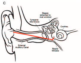
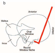
By correlating measurements from human cadaveric temporal bones with measurements from their respective CT scans, we defined key parameters necessary for designing an endoscope for intracochlear imaging using a minimally invasive approach through the external auditory canal.

Have a look at the images below.

image c.png

image b.png

Source: http://stankovic.hms.harvard.edu/fi...vic-md-phd/files/00129492-900000000-97335.pdf
 
@VRZ78 you will find the Texas TAMU Optical and Molecular Imaging lab site here: http://www.applegatelab.org/

Here is what they have to say about "understanding cochlear pathophysiology and function using picometer sensitive, spatially resolved vibrometry in the ear":

In humans, many of the diagnoses for hearing loss and vertigo or disequilibrium are based on a process of elimination, rather than positive proof of a particular pathology. This is especially true for diseases of the inner ear, which can lead to misdiagnosis and improper treatment.

You see what I mean?... "based on a process of elimination" and even "misdiagnosis and improper treatment". Audiology has played out its role!

Diseases of the inner ear while not life threatening can have a profound impact on the patient's quality of life.

Which includes tinnitus of course! Which is widely believed to be the result of inner ear damage. But of course we don't have proper tools to assess this damage or to do basic research and expand that body of knowledge about the ear and our hearing.

Hearing loss due to all causes affects over 30 million Americans. Conclusive diagnosis of the cause of hearing loss is not possible in most cases due to our inability to interrogate the soft tissues of the inner ear responsible for hearing. In fact, there is a fundamental lack of imaging technology capable of in vivo investigation of morphological and functional changes in the soft tissues of the inner ear of humans or animal models. This stems from the fact that inner ear in mammals is located deep inside the bone of the skull, the temporal bone in humans.

Yes, it's a big problem. But not many people are made aware of it. It's only about to get worse. When it becomes everyone's problem, I hope the "doctors" will have more tools and analysis to base their treatments on than just the audiograms.

Our work in this area is aimed at filling this void for both human patients and animal models of hearing disease. Our approach is fundamentally based on Optical Coherence Tomography (OCT), a medical imaging technique used clinically for imaging the eye, coronary arteries, and esophagus.

It doesn't say if they use micro OCT.

Our system development for animal imaging has focused on achieving extremely high sensitivity to vibration (~10 pm) at high imaging speeds using a fixture attached to a normal dissecting stereomicroscope.

This seems to suggest they are using some advanced version of OCT. Pico OCT? That's some insane level of precision.

Recent results include the first measures of tectorial membrane vibration within the unopened cochlea. For humans we are developing specialized systems that will enable imaging during surgery and awake patients in the clinic. This work is being done in collaboration with our collegues at Stanford University Department of Otolaryngology/Head and Neck Surgery.

In vivo, live view video stream of the patient's cochlea? Wow! This is the stuff I have been watching out for. Now, if they could only do it without making a hole in the ear drum... let's hope they just finish what they're up to right now and start pushing this technology out to the clinics. They can work on improvements later. This field is in bad need for a technology like this.

Sound is encoded within the auditory portion of the inner ear, the cochlea, after propagating down its length as a traveling wave. For over half a century, vibratory measurements to study cochlear traveling waves have been made using invasive approaches such as laser Doppler vibrometry. Although these studies have provided critical information regarding the nonlinear processes within the living cochlea that increase the amplitude of vibration and sharpen frequency tuning, the data have typically been limited to point measurements of basilar membrane vibration. In addition, opening the cochlea may alter its function and affect the findings. Here we describe volumetric optical coherence tomography vibrometry, a technique that overcomes these limitations by providing depth-resolved displacement measurements at 200 kHz inside a 3D volume of tissue with picometer sensitivity. We studied the mouse cochlea by imaging noninvasively through the surrounding bone to measure sound-induced vibrations of the sensory structures in vivo, and report, to our knowledge, the first measures of tectorial membrane vibration within the unopened cochlea.

https://www.ncbi.nlm.nih.gov/pubmed/25737536

I think it's safe to say that it's this study that has allowed for the recent discovery that the inner ear processes speech and music differently than other parts of the inner ear.

http://www.pnas.org/content/113/30/E4304.full

This is a study done by Linköping University, Oregon Health & Science University, Imperial College London and the Indian Institute of Technology.

It's nice to see that they are making progress and exchanging ideas. I am very hopeful that we might see something very useful come out of this in 2017.
 
Have we heard if they've done uOCT on a human?

Is staining required to get the colored uOCT images above or is that post image editing?

My suggestion to the Harvard team would be to start imaging live humans using the uOCT and try to perfect and that technique. This way they could serve as the diagnostician of for the Frequency/Decibel clinical trial. Aka do before and after photos. I'm sure Frequency would love that because a picture is worth a thousand word and with an independent team evaluating every person would be beating down their door for their compound. Heck even with the Boston traffic Harvard Frequency and Decibel are in the same city so it would be that hard to coordinate this.

Even if we had drugs that could effectively dampen the tinnitus sound or even eradicate it, who's to say we are making the right decision? Of course there are sufferers who can't stand the ringing. I would not blame them for wanting to take whatever magic pill we have to treat the symptom.

The magic pill/sound therapy will at best be a daily treatment. Taking a daily pill to dampen tinnitus likely will not make any future regenerative efforts any less successful. By daily meaning without it you'd revert back to the status quo ringing. The bigger risk with a daily pill would be the other health side effects induced by it see Trobalt.
 
Have we heard if they've done uOCT on a human?
They did that on "human cadaveric temporal bones". If that counts as "on a human"? See the earlier post.

Is staining required to get the colored uOCT images above or is that post image editing?
Yes, like most medical imaging techniques such as CT and MRI, the OCT devices normally produce grey-scale images. So, yes, staining is required to get some color on there. However, this is a proof of concept, so staining will not be done or even possible to do in patients.

No, this is not the result of Photoshop editing. If you click the link above you will see that the red color is rhodamine phallodin, green is neurofilament-H, and blue is Hoechst stain.

My suggestion to the Harvard team would be to start imaging live humans using the uOCT and try to perfect and that technique. This way they could serve as the diagnostician of for the Frequency/Decibel clinical trial. Aka do before and after photos.
It's not realistic that they will start using it as a diagnostic tool on humans yet. They have to make a hole in your ear drum and put the probe in. See the image above. While an ear drum can heal itself, you can't run away from the fact that it's an invasive procedure, even if they call it "minimally invasive".

They might be able to tweak the wavelength and energy output so that they don't have to make a hole in the ear drum. This may be easier said than done. It may not be possible to do that with OCT. There may also be legal limits to energy output. Perhaps more energy efficient electronic components will allow for more energy efficient OCT technology.

In the short term, this device will likely be used in with surgical procedures where you have to open the ear drum anyway. Therapeutic procedures like those being developed by Frequency will also benefit from this, because they aim to open the ear drum to deliver the drug.

Before and after comparison would be excellent for Frequency, Decibel, Auris, et. al. trials.

The magic pill/sound therapy will at best be a daily treatment. Taking a daily pill to dampen tinnitus likely will not make any future regenerative efforts any less successful. By daily meaning without it you'd revert back to the status quo ringing. The bigger risk with a daily pill would be the other health side effects induced by it see Trobalt.

I agree!
 
In the short term, this device will likely be used in with surgical procedures where you have to open the ear drum anyway.

Do you know why they don't use the same path that a nasal endoscope takes to reach the middle ear (and therefore, the cochlea)? I assume the NE reaches the middle ear via the Eustachian Tube.
 
I think at some point with the advanced technology it will be possible to reconnect the disconnected one via surgery
To have everyday utility it would have to be cheap, easy to use, usable in a clinic setting with minimal staffing and anaesthetic requirement, given what I anticipate the demand would be...so maybe in 50 years with (old) NASA-level funding. I would imagine that without meeting the above conditions any such development would have impossible criteria to be met before any treatment would get the OK, and...can you imagine how hard American Insurers would scramble to disallow payment for such a procedure?
 
Do you know why they don't use the same path that a nasal endoscope takes to reach the middle ear (and therefore, the cochlea)? I assume the NE reaches the middle ear via the Eustachian Tube.
The angle is wrong to access the RWM. Given the small space, I don't know that there is a way around this.
 
The angle is wrong to access the RWM. Given the small space, I don't know that there is a way around this.

They are able to do a full stapedotomy with a nasal endoscope, though. Is the RWM that far off from it? I thought they were next to each other. Maybe the OCT hardware has a specific shape that can't take bends.


Log in or register to get the full forum benefits!

Register

Register on Tinnitus Talk for free!

Register Now