The article on Eurekalert was not clear about what that previous research was that identified the first cause of hidden hearing loss (HHL). I thought they were referring to a previous work by Corfas at Michigan university. I now realize that they were referring to the work by Liberman at Massachusetts Eye and Ear. I thought it seemed familiar...
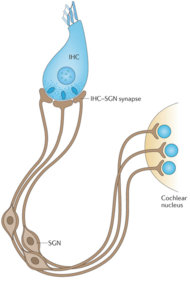
HHL has been proposed to lead to hyperactivation of central auditory synapses, thus possibly contributing to tinnitus. Until now, loss or dysfunction of IHC synapses has been the only proposed mechanism of HHL.

You can see the location of IHC synapses in the image above.
We found that acute Schwann cell loss causes rapid auditory nerve demyelination, which is followed by robust Schwann cell regeneration and axonal remyelination. Surprisingly, this transient Schwann cell loss results in a permanent auditory impairment characteristic of HHL.
In other words the Schwann cell loss and axonal demyelination is not permanent (hence "transient"). But the natural restoration that follows is not perfect. Myelin is a protective sheath that surrounds nerve fibers.
However, this HHL differs from that seen after noise exposure or ageing in that it occurs without alterations in synaptic density but rather correlates with a specific and permanent disruptions of the first heminodes at the auditory nerve axon close to the IHCs.
In other words there is no nerve fibre loss. There is a disruption in the Heminode which sits at the Habenula perforata. Heminode is a node of Ranvier situated at the junction of myelinated (protected) axons and bare (unprotected) axons. In the cochlea this happens at a place called Habenula perforata.
Furthermore, the two types of HHL occur independently (noise exposures that cause HHL do not disrupt heminodes) and they are additive. Together, these results uncover a new mechanism for the pathogenesis of HHL and a new consequence of myelin defects on the normal function of the nervous system.
In other words myelin defects are bad, there are two types or causes for HHL, and they can add up.
To help you navigate around and better understand this I have include two images below.
Location of Habenula perforata:
Schematic view of cochlear nerve fibres:
Explanation:
Schematic illustration of the peripheral glial cells in the adult human cochlea. Satellite glial cells (green) envelop all spiral ganglion neuron (SGN) cell bodies. Non-myelinating Schwann cells (light blue) ensheath both the central and peripheral processes of the type II SGNs (yellow) that innervate the outer hair cells (OHC). Myelinating Schwann cells (dark blue) ensheath and myelinate both processes of the type I SGNs (red) that innervate the inner hair cells (IHC). Beyond the habenula perforata, in the organ of Corti, neither Schwann cell types ensheath the most distal part of the peripheral processes of type I and type II SGNs.

 Manager
 Member